Thursday, September 7, 2023

Multiple Focal Choroidal Excavations in Association with Protein Rich Diet


Introduction

Choroidal excavation is a novel entity that is diagnosed with optical coherence tomography (OCT). In 1959, Klien, et al. [1] first described a concave-shaped chorioretinal abnormality with undifferentiated choriocapillaris and retinal pigment epithelium. Then in 2006, Jampol [2] defined similar changes with OCT and first used the term of focal choroidal excavation (FCE). In 2011, Margolis, et al. [3] described two FCE patterns, based on the shape of the lesions: nonconforming, if the photoreceptors are detached from retina pigment epithelium (RPE) and conforming, when the RPE follows the photoreceptor layer without any optically clear space between the outer retina and RPE. These two forms are also thought to convert to each other during the clinical process of the disease [4]. Although it is known to be mostly idiopathic, its relationship with central serous chorioretinopathy, choroidal neovascular membrane and inflammatory diseases such as multiple evanescent white dot syndrome and Vogt Koyanagi Harada disease has been reported [5]. In this report, we present a patient with multiple focal choroidal excavation following protein-rich diet.

Case

21-year-old male patient applied with blurred vision in his right eye since one week. He was on a protein rich diet since he was doing bodybuilding and had been using approximately 2g/kg/day protein powder since 3 months. His best corrected visual acuity was 0,6 in his right eye (RE) and 0,9 in his left eye (LE). There was no refractive disorder. Slit lamp examination and intraocular pressure were normal. Fundus examination revealed cavitary lesions in the macula of the RE (Figure 1a) and pigmentary changes above the macula of the LE (Figure 1b). OCT showed multiple conforming (Figure 2a) and non-conforming (Figure 2b) type of focal choroidal excavations with small retinal pigment epithelium detachments in the right eye and a small retinal pigment epithelium detachment in the left eye (Figure 2c). When evaluated with fundus autofluorescence (FAF), there have been hypo and hyperautofluorescent areas in the macula of the right eye in accordance with the pigmentary changes (Figure 3a) and hypoautofluorescent area above the macula of the left eye (Figure 3b). Fundus fluorescein angiography showed multiple hyperfluorescent window defects in the macula of the RE and hyperfluorecence consistent with window defect above macula of the LE. There was no ischemia, leakage, macular edema and all vascular structures were normal . Protein powder stopped and the patient was examined 6 months after the diagnosis. No changes in the findings were detected.


Read more about this article : https://lupinepublishers.com/ophthalmology-journal/fulltext/multiple-focal-choroidal-excavations-in-association-with-protein-rich-diet.ID.000158.php

Read More About Lupine Google Scholar:    https://scholar.google.com/citations?view_op=view_citation&hl=en&user=T4c9RDkAAAAJ&citation_for_view=T4c9RDkAAAAJ:blknAaTinKkC


Friday, August 25, 2023

GLS-1027 to Treat Anterior Uveitis in a Cavalier King Charles Spaniel Dog

 
Abstract
Non-infectious, autoimmune anterior uveitis is a significant cause of ocular disease in both dogs, horses, and humans with the potential for significant vision loss. The mainstay of treatment is topical and/or systemic corticosteroids, however, animals, similar to humans, are not uncommonly resistant to therapy. Additionally, prolonged corticosteroid treatment may cause systemic and ocular toxicity. We describe here a case of steroid-resistant uveitis successfully treated with GLS-1027, an immunomodulatory compound that inhibits Th17 maturation and release of multiple inflammatory cytokines involved in the pathogenesis of uveitis. GLS-1027 may provide an alternative as a steroid-sparing therapeutic option.

Introduction
Non-infectious, anterior uveitis is a significant cause of ocular disease in dogs, horses, humans, and other species that has the potential to lead to vision loss and blindness. While the disease is most commonly considered as autoimmune for humans and the most common etiology in dogs [1,2] infectious disease neoplastic etiologies must also be considered [2,3]. In all species, the primary goal of treatment is sight preservation with secondary goals to limit pain, discomfort, photophobia, and in dogs minimizing secondary sequelae such as posterior synechia [3]. In animals, topical corticosteroid treatment combined with topical mydriatics with or without the use of systemic anti-inflammatory medications represents the standard of care for anterior uveitis [2,4]. While immunosuppressives are considered the standard for steroidresistant disease in humans [5], these are seldom used in animals due to toxicity and cost. Studies in animals have reported that approximately 1-35% of the total topical corticosteroid dose is systemically absorbed, [4] and may result in systemic side effects, including endocrinopathies [3]. Steroid-induced ocular pathology, including the development of glaucoma, cataract formation, and keratopathy are not uncommon in dogs with prolonged corticosteroid use [4] with a similar side effect profile for humans. Therefore, a safe and effective alternative to corticosteroid treatment for chronic recurrent idiopathic uveitis is warranted. Autoimmune uveitis in humans [6] and horses [7,8] has been attributed to Th17 mediated pathology with a complex interplay between a number of inflammatory cytokines including TNFα and IL-6. GLS-1027, [S,R]-3-phenyl-4,5-dihydro-isoxazoleacetic acid, is a small molecule compound with anti-inflammatory activity currently in clinical development. GLS-1027 is a potent inhibitor of lipopolysaccharide (LPS)-induced inflammation characterized by normalization of levels of the cytokines tumor necrosis factor (TNFα), interleukin (IL) 1β, and IL-6 both in vitro and in murine animal models [9-12]. Additional unpublished data has shown that GLS-1027 inhibits IL-17 and IL-23 mediated activation and downregulates Th17 T cells.

In this paper, we describe a case report of the use of GLS-1027 to treat canine uveitis.


Read More About Lupine Google Scholar: https://scholar.google.com/citations?view_op=view_citation&hl=en&user=T4c9RDkAAAAJ&citation_for_view=T4c9RDkAAAAJ:JV2RwH3_ST0C


Tuesday, August 15, 2023

Cauchy Modeling as a New Trend in Research of Fullerene- Type Nanomaterials


Abstract

In the work, based on the analysis of heat and gas with acoustic phonons by fullerene, the physical criterion Cauchy is the kind of thermal strength before spring strain, which is caused by the nanotube fullerene

Keywords: Thermal Conductivity; Fullerene; Optimization; Numbers Similarity Numbers

Introduction

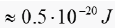
Studies of the thermal conductivity of structured nano systems draw attention to the fact that in micro- and nanoelectronics the urgent task of heat dissipation released during the operation of electrical elements [1-6]. So, in the process of electrical switching of the element energy is consumed Lupinepublishers-openaccess-ophthalmology-journal which is on the verge of thermal fluctuations Lupinepublishers-openaccess-ophthalmology-journal This means that a billion (109) transistor chip uses order energy in a single act 10-3J. Then, in order of speed 1GHz , the energy consumed can reach values Lupinepublishers-openaccess-ophthalmology-journal far exceeding the power of a jogging electric kettle Lupinepublishers-openaccess-ophthalmology-journal

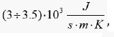
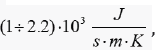
In order to remove Joule-Lenz heat in electric nanoelements, both microchannels with liquid carriers of heat and conductive elements, such as nanocomposite ceramics filled with carbon nanotubes, can be used, which, depending on their chiral characteristics, can alter the physical properties of nano cells. Thus, it was established [9-12] and confirmed by theoretical estimates [13,14] that the thermal conductivity at room temperature of a carbon nanotube can reach values of Lupinepublishers-openaccess-ophthalmology-journal which significantly exceeds the thermal conductivity of diamon Lupinepublishers-openaccess-ophthalmology-journal which was considered one of the best heat conductors. Carbon nanotubes are thermally stable, characterized by high electrical conductivity [10], high electron mobility [11], and a large specific surface value [15]. It is believed [16] that the abnormally high thermal conductivity of carbon nanotubes is due to their regular structure and the small number of defects and impurity centers in them. In the elementary cell of graphene there are two carbon atoms, so the dispersion spectrum consists of three optical and three acoustic branches, among which the longitudinal and transverse acoustic modes correspond to the speed of sound 2130 and 1360 M/s


Read more about this article : https://lupinepublishers.com/ophthalmology-journal/fulltext/cauchy-modeling-as-a-new-trend-in-research-of-fullerene-type-nanomaterials.ID.000156.php

Read More About Lupine Google Scholar: https://scholar.google.com/citations?view_op=view_citation&hl=en&user=T4c9RDkAAAAJ&citation_for_view=T4c9RDkAAAAJ:M3NEmzRMIkIC

Thursday, July 27, 2023

A Review of Metabolic Sensors in Glaucoma


Abstract

Glaucoma is the second leading cause of irreversible blindness worldwide. It is a multifactorial, progressive, chronic optic neuropathy that is characterized by loss of retinal ganglion cells (RGC) and optic nerve head (ONH) cupping including extra cellular matrix (ECM) remodelling and fibrosis at the lamina cribrosa (LC). Clinically this results in chronic, progressive peripheral visual field loss. The pathogenesis of glaucoma is not yet fully understood. Therefore, there is an urgent need to identify and target the underlying mechanisms governing ECM remodelling of the LC, in order to stop the progressive, chronic damage to the LC/ONH and irreversible visual field loss. This review identifies and examines some of the key metabolic processes and cellular sensors involved in the pathogenesis of ECM fibrosis in general but herein specifically in glaucoma, including mitochondrial dysfunction and adenosine monophosphate activated protein kinase (AMPK) upregulation. Furthermore, the development of novel therapeutics such as nicotinamide (NAM) and metformin are discussed as promising potential future therapeutic options for glaucoma.

Read more about this article : https://lupinepublishers.com/ophthalmology-journal/fulltext/a-review-of-metabolic-sensors-in-glaucoma.ID.000155.php

Read More About Lupine Google Scholar: https://scholar.google.com/citations?view_op=view_citation&hl=en&user=T4c9RDkAAAAJ&citation_for_view=T4c9RDkAAAAJ:maZDTaKrznsC

Saturday, July 8, 2023

A Review of Achromatopsia


Abstract

Congenital achromatopsia is a hereditary form of day blindness caused by cone photoreceptor dysfunction, with an incidence of approximately 1 in 30,000. This inherited disorder is characterized by a lack of color discrimination, nystagmus, photophobia, and low visual acuity (< 0.2). The most typical genetic mutations are autosomal recessive changes in CNGA3, CNGB3, GNAT2, PDE6H, PDE6C, or ATF6. It should not be confused with cerebral achromatopsia, which is an acquired form of total color blindness that can result from illness, trauma, or some other cause. Color plays an essential role in our lives. It can change actions, influence thinking, and cause reactions. As a powerful type of communication, color is irreplaceable. Green means “go” and red means “stop”. Traffic lights send this global meaning. Color vision deficiency, creates challenges in the daily lives of those who have an insufficient visual sense.

Purpose: The aim of this review is to examine the literature published on achromatopsia and summarize the diagnosis, management, genetic characteristics, and the recent advances in gene therapy.

Conclusions: Congenital achromatopsia is a complex inherited disease. Management of achromatopsia is multifaceted. There is currently no cure for achromatopsia, although gene therapy is a therapeutic option already being studied in clinical trials. The most recent study in human adults can be classified as safe and positive in terms of efficacy.

Keywords: Achromatopsia (ACHM); Gene therapy

Introduction

Achromatopsia is a condition characterized by a partial or total absence of color vision [1]. People with complete achromatopsia cannot perceive any colors; they only see white, black, and shades of gray. Incomplete achromatopsia is a milder form of the disease that allows some color discrimination [1]. Oliver Sacks, in The Case of Color-blind Painter [2], transcribe the disturbing story of one of his cerebral achromatopsia patients after a car accident in the following way: Mr. I. could hardly tolerate the changed aspects of people (“like animated grey statues”) any more than he could tolerate his own appearance in the mirror: he shunned social intercourse and found sexual intercourse impossible. He saw people’s fesh, his wife’s fesh, his own fesh, as an abhorrent grey; “fesh-colored” now appeared “rat-colored” to him.


Read more about this article : https://lupinepublishers.com/ophthalmology-journal/fulltext/a-review-of-achromatopsia.ID.000154.php


Read More About Lupine Google Scholar: https://scholar.google.com/citations?view_op=view_citation&hl=en&user=T4c9RDkAAAAJ&citation_for_view=T4c9RDkAAAAJ:k_IJM867U9cC

Tuesday, June 27, 2023

Intravitreal Injection of Ranibizumab in Macular Edema Secondary to Retinal Vein Occlusion


Abstract

Aim: This study aimed to evaluate the safety and efficacy of intravitreal Ranibizumab 0.5mg in the treatment of macular edema secondary to retinal vein occlusion.

Patients & Methods: This was a prospective interventional analytical study included 39 eyes of 39 patients with retinal vein occlusion. Ophthalmic examination included assessment of visual acuity, measurement of intraocular pressure, and fundus examination. All patients were scanned using Swept source optical coherence tomography (3D DRI OCT Triton [plus], Topcon Corporation, Tokyo, Japan) to assess central macular thickness. The changes of visual acuity, IOP, and central macular thickness were assessed. Data were analyzed via Kolmogorov-Smirnov test and Wilcoxon signed rank.

Results: The mean age was 56.56 ± 9.6, 48.7% were male and 51.3% were females. Hypertension was detected in 69.2%, and hyperlipidemia in 2.6%. The mean best corrected visual acuity was 1.5 logMAR, 1.00 logMAR,1.00 logMAR, preoperative, fourth month, six months postoperative, respectively, (p<0.001). The mean central macular thickness was 675 μ, 306 u, 264 u, preoperative, fourth month, six months postoperative, respectively, (p< 0.001). The OP was 16.5 mmHg, 16.9 mmHg, 17.1 mmHg, preoperative, fourth month, six months postoperative, respectively, (p=0.423). There were no observed significant ocular adverse events such as ocular inflammation, sterile and infectious endophthalmitis, or sustained increase in intraocular pressure with the use of intravitreal ranizumab injections.

Conclusion: Intravitreal Ranibizumab injections as monotherapy have shown promising results with BCVA improvement and a decrease of central macular thickness in patients with macular edema secondary to retinal vein occlusion.

Keywords: Ranibizumab; Macular Thickness; OCT; Visual Acuity

Introduction

Retinal vein occlusion (RVO) is the most common retinal vascular disease after diabetic retinopathy [1]. Depending on the area of retinal venous drainage effectively occluded it is broadly classified as either central retinal vein occlusion (CRVO), hemispheric retinal vein occlusion , or branch retinal vein occlusion (BRVO) [2]. Although the exact etiology of RVO remains elusive, it is likely to follow a thrombotic event. In CRVO this may occur in the central retinal vein (CRV) at the lamina cribrosa or at a variable distance in its journey within the optic nerve posterior to the lamina cribrosa [2]. Hypoxia-induced expression of vascular endothelial growth factor (VEGF) is thought to be a trigger for macular edema. High intravitreal levels of VEGF have been found in patients with retinal vein occlusion [3]. Upregulation of VEGF is associated with breakdown of the blood-retina barrier with increased vascular permeability resulting in retinal edema, stimulation of endothelial cell growth, and neovascularization [4,5]. Macular edema leads to vision loss in many patients with either central or branch retinal vein occlusions (CRVO or BRVO). BRVO is the more common of the two presentations, accounting for approximately 80% of RVO [6].
Recently, there has been interest in the use of vascular endothelial growth factor (VEGF) inhibition in the treatment of RVO because of the observation of increased VEGF in the vitreous and aqueous of patients with these conditions [7].

Read more about this article : https://lupinepublishers.com/ophthalmology-journal/fulltext/intravitreal-injection-of-ranibizumab-in-macular-edema-secondary-to-retinal-vein-occlusion.ID.000153.php


Read More About Lupine Google Scholar: https://scholar.google.com/citations?view_op=view_citation&hl=en&user=T4c9RDkAAAAJ&cstart=20&pagesize=80&citation_for_view=T4c9RDkAAAAJ:isC4tDSrTZIC

Tuesday, June 13, 2023

Visual Impairment and its Rehabilitation: A Review


Abstract

Visual impairment (VI) is a condition of reduced visual performance that cannot be remedied by refractive correction (spectacles or contact lenses), surgery or medical methods. Visual Impairment is a broader term that encompasses both Low vision and Blindness. The prevalence of visual impairment is estimated to be around 2.2 billion in the world as per World Health Organization (WHO). Causes of Visual Impairment differs significantly in different regions. Saying that, still cataract and refractive errors are main causes of visual impairment globally. Owing to its negative impact on the individual’s dailies, professional life, productive time, confidence, and self-respect, VI needs to be addressed timely and appropriately. Visual impairments do have profound consequences for the individual, however effective rehabilitation services can restore independence and therefore ameliorate these consequences and restore the individual’s quality of life.

Keywords: Visual Impairment; Blindness; low Vision; Rehabilitation; Optical Devices; Non-Optical Devices

Introduction

Impairment of any kind (sensorial or motor) is a bane for any human being. With visual impairment being one of the leading causes of sensorial impairment, it is necessary to acknowledge its prevalence, causes and methods to avoid them, if possible. Also, at the same time, it is important to understand that the term “visual impairment”, “blindness” and “Low vision” are not synonymous; rather visual impairment includes low vision as well as blindness. Simply, Visual impairment (VI) is a condition of reduced visual performance that cannot be remedied by refractive correction (spectacles or contact lenses), surgery or medical methods [1]. Various definitions of visual impairment have been given by various concerned authorities. International Statistical Classification of Diseases and Related Health Problems (ICD) defines visual impairment categories primarily on the basis of recommendations made by a World Health Organization (WHO) Study Group in 1972 [2] and defines as:

• low vision is defined as visual acuity of less than 6/18, but equal to or better than 3/60, or a corresponding visual field loss to less than 20 degrees in the better eye with best possible correction
• blindness is defined as visual acuity of less than 3/60, or a corresponding visual field loss to less than 10 degrees in the better eye with best possible correction.


Read more about this article : https://lupinepublishers.com/ophthalmology-journal/fulltext/visual-impairment-and-its-rehabilitation-a-review.ID.000152.php


Read More About Lupine Google Scholar: https://scholar.google.com/citations?view_op=view_citation&hl=en&user=T4c9RDkAAAAJ&citation_for_view=T4c9RDkAAAAJ:TFP_iSt0sucC

Monday, April 10, 2023

Headache and Eye-A Mutual Relationship


Opinion

Headache is probably the commonest neurological complaint worldwide. According to the World Health Organization (WHO), 1.7-4% of the adult population of the world have headaches on 15 or more days every month. Sometimes these symptoms are misunderstood and confused with Migraine Attacks or with normal tension headaches. It is also one of the commonest problems because of which people come for a glass/eye check-up. Very often the belief is that a change of eye power is the reason for headaches. Students who constantly keep reading books or keep doing their academic exercises have more chances to suffer from eye related problems like Eye strain, Fatigue, Eye pain, Headache etc. When a patient comes to you with complaints like headache after some continuous reading it is important to be able to distinguish if it is related to the eyes or not. Headache can be felt in a variety of places on the head, including the temples, sinuses, forehead and back of the head. Eye pain associated with a headache can be caused by a few different types of headaches, as well as other seemingly unrelated conditions. But regardless of the cause, this type of pain can be mild, moderate, or even unbearable.


Read more about this article : https://lupinepublishers.com/ophthalmology-journal/fulltext/headache-and-eye-a-mutual-relationship.ID.000151.php


Read More About Lupine Google Scholar: https://scholar.google.com/citations?view_op=view_citation&hl=en&user=T4c9RDkAAAAJ&citation_for_view=T4c9RDkAAAAJ:bEWYMUwI8FkC

Wednesday, March 29, 2023

Effects of Long Term Use of Prostaglandin Analogues with and Without Preservative on Normal Conjunctival Flora of Glaucoma Patients


Abstract

Purpose: The effects of long term use of Benzalkonium chloride-preserved prostaglandin analogue eye drop on conjunctival bacterial flora was investigated and compared with preservative-free prostaglandin analogue eye drop

Methods: Conjunctival swabs were collected from 48 patients of glaucoma who had instilled either BAK-preserved latanoprost or preservative-free travoprost eye drops for 1 year. The bacterial characteristics and sensitivity patterns of the bacterial isolates from the conjunctival swabs of the two groups were compared between each other and also with conjunctival bacterial flora from normal healthy controls.

Results: There was no significant difference in culture positivity among the three groups (p = 0.99). 100% (13/13) isolates from Latanoprost group were Gram positive organisms. In the Travoprost group, 12 out of 14 isolates (85.7%) from 13 eyes were gram positive while the rest 2 bacteria (14.3%) were gram negative. In Controls, 14 out of 16 (87.5%) isolates were gram positive and the rest were gram negative. Methicillin-resistance was seen in 72.7% (8/11) of the CONS isolated from Latanoprost group, while 45.5% (5/11) of the CONS isolated from preservative free travoprost group were Methicillin resistant. While in the controls 45.4% (5/11) of the isolated CONS were Methicillin resistant. The occurrence of Methicillin resistant CONS (MR CONS) was higher in latanoprost group than the other two groups, though not statistically significant. Resistance to commonly used antibiotics was comparatively higher in BAK-preserved latanoprost group. Not a single gram positive bacteria was resistant to Vancomycin

Conclusion: Long-term use of prostaglandin analogues for glaucoma may adversely affect the antibiotic-resistance of indigenous bacterial flora of the eye by increasing the resistance against common antibiotics including Methicillin resistance.

Read more about this article : https://lupinepublishers.com/ophthalmology-journal/fulltext/effects-of-long-term-use-of-prostaglandin-analogues-with-and-without-preservative-on-normal-conjunctival-flora-of-glaucoma-patients.ID.000150.php


Read More About Lupine Google Scholar: https://scholar.google.com/citations?view_op=view_citation&hl=en&user=T4c9RDkAAAAJ&citation_for_view=T4c9RDkAAAAJ:iH-uZ7U-co4C

Monday, March 20, 2023

Anti-Oxidant Effect of Lycium Barbarum Polysaccharides on the Retina of Streptozotocin-induced Diabetes Rats


Abstract

Aim: To investigate the effect of Lycium barbarum polysaccharides (LBP) on the oxidation damage of retina of streptozotocininduced diabetes rats.

Methods: The diabetes rats were induced by injection streptozotocin intraperitoneally. LBP was intragastrically administered to the rats of the LBP group. Body weight and blood glucose were measured every four weeks. In the 24th week, the SOD and MDA of retina were measured, the expression of VEGF mRNA of retina was tested by RT-PCR, the ultrastructure was observed by transmission electron microscopy.

Results: The body weight decreased and the blood glucose increased in DM group and LBP group (P<0.01, VS control group). The body weight and blood glucose is the same level between DM group and LBP group (body weight P=0.503, blood glucose P=0.984). The SOD in DM group decreased and the MDA increased (P<0.05, VS control group). The SOD in LBP group increased and the MDA decreased (P<0.05, VS DM group). The expression of VEGF mRNA was highest in DM group and lowest in control group, and decreased in LBP group (P<0.05, VS DM group). The ultrastructure changes occur in almost all retinal nerve tissues in DM group, as represented by the number and morphology of mitochondria. A small amount of mild mitochondrial changes could be observed in the endochylema of bipolar and Müller cells in LBP group.

Conclusion: LBP can reduce the oxidative damage, alleviate the pathological changes in mitochondria, prevent the apoptosis of nerve cells and block the progression of diseases to the vascular tissue.

Keywords: Lycium Barbarum Polysaccharides; Diabetes; Anti-Oxidant; Diabetic Retinopathy

Thursday, March 9, 2023


Abstract

Purpose: To evaluate the visual performance after bilateral implantation of an extended depth of focus (EDOF) intraocular lens (IOL).

Methods: In this multicenter, prospective, observational study, the postoperative outcomes of 96 patients who underwent bilateral cataract surgery with EDOF IOL (TECNIS Symfony) were assessed.

Results: Mean decimal visual acuity results showed a binocular corrected distance visual acuity (CDVA) of 1.10 ± 0.18, uncorrected distance visual acuity (UDVA) of 1.04 ± 0.17, uncorrected intermediate visual acuity (UIVA) of 0.96 ± 0.16, and uncorrected near visual acuity (UNVA) of 0.68 ± 0.18. Overall, 76% of the patients achieved spectacle independence across all distances, and more than 85% reported no or mild dysphotoptic phenomena. On a scale from 0 to 10, median patient satisfaction score was 9 for far, 9.5 for intermediate, and 8 for near vision.

Conclusions: The Symfony EDOF IOL provided excellent distance and intermediate visual outcome and functional near visual acuity. The visual results were associated with high levels of spectacle independence and patient satisfaction.


Monday, February 27, 2023

An Ophthalmologist look at Art

Introduction

In the 1990s Dr. María Dolores Cortés lent me a very interesting book called “An Ophthalmologist look at art” written by Arthur Linksz M.D. The author makes several analyses, observations and conjectures about the portraits of people who accepted the indications of the painter, although there were some important persons who wanted to impose his desires and that created problems for the painter; then it’s about self-portraits, some made in front of a mirror and others taken from a photograph; later it deals with the direction of reading and writing, from left to right in the western and right to left in the East, and ends with uncapítulo on the commented astigmatism of the Greco. I think an ophthalmologist who likes art, especially painting should have read it. In short, when referring to portraits, he points out that people receive the light on the left side so as not to cover it if the painter is right-handed, except when an important person did not meet the painter’s demands.


Friday, February 17, 2023

Thymoquinone, Lipids and Herbal Medication


Opinion

Georges Lemaitre’s first-time atom, or the big bang in English, was the beginning of the universe. From a very small thing began to appear the elements, matter, gases, liquids, like water, then the nebulae, within them the solar systems with their planets, satellites and comets. A small solar system in a corner of a small galaxy called the Milky Way had several planets, some large and other boys; among the little ones one had water where over time surfaces appeared with chlorine arrow that needed the sun’s rays to live and develop, then began to form beings that could move, the animals of which there were microscopic and later reached gigantic. Many millennia later Eve and Adam appeared, who were very good animals but one day they came up with sin and they stopped being animals. The animals did not sin, and first time felt naked. If Eve had not sinned today, we would all walk naked, we would be very good little animals and Jesus of Nazareth would not have had to come, because He came for sinners, not for the saints or the virtuous. A good ateor, sincerely, you can say that you do not believe in God, you must believe in the primal atom, which by analyzing its existence can be considered to be God.


Read more about this article : https://lupinepublishers.com/ophthalmology-journal/fulltext/and-later-they-came-to-the-primigenio.ID.000146.php


Read more about Lupine Google Scholar: https://scholar.google.com/citations?view_op=view_citation&hl=en&user=T4c9RDkAAAAJ&citation_for_view=T4c9RDkAAAAJ:4JMBOYKVnBMC

Wednesday, February 8, 2023

Thymoquinone, Lipids and Herbal Medication


Abstract

Allopathic drugs used as hypolipidemic agents have number of unwanted effects. Herbal therapy for Hyperlipidemia is getting attention due to their less frequent side effects. In this study we have compared hypolipidemic effects of Fenofibrate 40 mg with Nigella sativa. Seventy-five hyperlipidemic patients from National Hospital Lahore were enrolled for study. Consent was taken from all enrolled participants and were divided in three equal numbers i.e. twenty-five in each group. Group 1 was on Nigella sativa, group 2 was on Gemfibrozil and third group was on placebo therapy. They were advised to take drugs for two months. After completion of study pretreatment and post treatment values of LDL cholesterol were analyzed statistically. In 25 patients who were on Nigella sativa, their LDL cholesterol decreased from 191.14±3.45 to 159.40±2.98 mg/dl. 31.7 mg/dl LDL reduction was observed when compared with placebo group. In 25 patients who were on Fenofibrate 40 mg, their LDL cholesterol decreased from 197.77±3.91 mg/dl to 159.62±2.20 mg/dl. LDL reduction in this group was 38.2 mg/dl. These changes are highly significant with p-values of <0.001. We concluded from this study that hypolipidemic characteristic of Nigella sativa is comparable and therapeutically as effective as traditionally used hypolipidemic medication Fenofibrate.

Read more about this article : https://lupinepublishers.com/ophthalmology-journal/fulltext/thymoquinone-lipids-and-herbal-medication.ID.000145.php

Read more about Lupine Google Scholar: https://scholar.google.com/citations?view_op=view_citation&hl=en&user=T4c9RDkAAAAJ&citation_for_view=T4c9RDkAAAAJ:_Qo2XoVZTnwC

Friday, January 6, 2023

Pattern of Congenital Ocular Anomalies among Children Seen at a West African Tertiary Eye Care Centre


Abstract

Purpose: To describe the pattern of presentation of congenital ocular anomalies among children seen at Department of Ophthalmology (Guinness Eye Centre), Lagos University Teaching Hospital, Lagos, Nigeria.

Methods: A retrospective chart review of children below the age of 16 years who were diagnosed of any type of congenital ocular anomaly at the Pediatric Ophthalmology Clinic of Lagos University Teaching Hospital (LUTH) between January, 2012 and December, 2018 was done. Information concerning age at presentation, gender, affected eye(s), visual acuity and type of congenital anomaly were retrieved from the case files.

Results: Seven hundred and forty-five eyes of 470 patients with congenital anomalies which constituted 13.6% of all the new pediatric ophthalmic consultations were studied. Two hundred and seventy-five (58.5%) children had bilateral ocular involvement while 262 (55.7%) presented within the first year of life. The median age was 0.92 years with an interquartile range of 2.67 years. There were 255 (54.5%) males with a male to female ratio of 1.2:1. Congenital cataract was the most common congenital ocular anomaly documented in 224 (30.1%) eyes of 133 patients. This was followed by congenital squint (131 eyes, 17.6%), congenital glaucoma (91 eyes, 12.2%) and corneal opacity (52 eyes, 7.0%). Overall, cataract, squint, glaucoma, corneal opacity, nasolacrimal duct obstruction and ptosis accounted for 79.0% of the congenital ocular anomalies documented in this study.

Conclusion: Congenital ocular anomalies accounted for 13.6% of Paediatric ophthalmic consultations in this study. Congenital cataract, squint, glaucoma, corneal opacity, nasolacrimal duct obstruction and ptosis were the most common congenital ocular anomalies observed.


Read more about this article : https://lupinepublishers.com/ophthalmology-journal/fulltext/pattern-of-congenital-ocular-anomalies-among-children-seen-at-a-west-african-tertiary-eye-care-centre.ID.000144.php

Read more about Lupine Google Scholar: https://scholar.google.com/citations?view_op=view_citation&hl=en&user=T4c9RDkAAAAJ&citation_for_view=T4c9RDkAAAAJ:e5wmG9Sq2KIC

Wednesday, December 14, 2022

Acute Corneal Hydrops in a Young Patient with Down Syndrome


Abstract

A case of severe acute corneal hydrops with keratoconus associated with Down syndrome described in a 16-year-old male patient presented with a sudden unilateral painful visual loss in his left eye. The anterior segment of his left eye showed a severe diffuse edematous ectasia of acute corneal hydrops. Eye rubbing of his left eye was observed. Continued gradual improvement in his visual acuity and a central corneal scar were detected.

Introduction

Down’s syndrome is frequently associated with keratoconus, a chronic non-inflammatory disorder which is characterized by progressive thinning of the cornea leading to scarring and progressive stromal thinning. Its incidence in Down’s syndrome has been reported up to 15% [1]. Acute corneal hydrops is characterized by marked stromal edema due to a tear in Descemet membrane (DM) which permits aqueous to enter the corneal stroma and epithelium [2-3].

Read more about this article : https://lupinepublishers.com/ophthalmology-journal/fulltext/acute-corneal-hydrops-in-a-young-patient-with-down-syndrome.ID.000143.php


Read more about Lupine Google Scholar: https://scholar.google.com/citations?view_op=view_citation&hl=en&user=T4c9RDkAAAAJ&citation_for_view=T4c9RDkAAAAJ:R3hNpaxXUhUC

Friday, June 24, 2022

Lupine Publish | Laser Clot Blast Therapy for Central Retinal Venous Occlusion

Lupine Publishers | Trends in Ophthalmology Open Access Journal 
 

Abstract

Purpose: To report Yag laser clot blast therapy for central retinal venous occlusion.

Methods & Materials: Prospective study of 10 patients. Best visual acuity was finger counting at 1 meter in all the cases, which were subjected to the treatment. The cases were followed up for 2-4 years (2.5 years).

Results: In 10 cases visual acuity improved to 0.4 (20/50) in 1st week treatment. The best final visual acuity improvement was 20/30 in 8 and 20/40 in 2 cases.

Conclusions: Early Yag laser clot blast therapy is useful for rapid improvement of the visual acuity and prevents the complications secondary to central venous occlusion.

Most of the cases of the occlusion of central retinal vein invariably turned out to be unsuccessful with medical therapy given in the form of fibrinolytic agents, anticoagulants, hemodilution” and steroid4. The cases that present with Ischemic central retinal venous occlusion with visual acuity of 20/200 or less are typically unfavorable’. In most instances, even the laser photocoagulation does not yield a visual benefit compared with the natural course of the diseases. Surgical decompression of branch retinal vein occlusion via arteriovenous crossing sheathotomy has been tried successfully by some67’8 to treat these cases but had led to complications associated with interventional vitrectomy including accelerated nuclear sclerotic cataract formation and retinal detachment. Others9 have tried to develop chorioretinal anastomosis at the site of the venous block. However, others1° are trying to cannulise the blocked central retinal vein. As no single treatment for central retinal vein is yet successful, the authors tried a new modality of treatment in cases of central venous retinal occlusion.

Methods and Materials

The experimental procedure of Yag laser clot blast therapy was done in two blind eyes due to old optic atrophy after taking a written consent. There were no complications seen at time of laser application in the post laser follow up of 12 months. After this the present study was conducted at G.G.S.I.E Research & Cure Centre from 1996 to 2001 after taking written consent of the patients. There were 10 cases of central retinal venous block of ischemic type. The ages of patients ranged from 24 to 70 years. In all the cases, best visual acuity was recorded with Snellen projector chart [1]. Charting of fields and ophthalmoscopic examination were done. The eyes were subjected to retinal fluorescein angiography. The patient was made to sit on a slit lamp. After insertion of mainster pan fundus contact lens, the Yag laser bursts of 1 .0milli Joules were started on the vein where it was entering as a branch or main trunk on the optic disc, the settings of the laser were kept at 125micron anterior. If the energy released was less, it was increased to 2 to 3milli Joules. After giving 4 bursts at the disc, a total of 6-12 bursts were given along the entire length of the blocked vein and venules till the 2nd A.V. crossing (Figure1 ). The patients were sent back and were asked to report once in a week till 4th week and then fortnightly till a follow up of 2-4 yrs (2.5yrs) [2]. A complete examination of the eye including recording of best visual acuity, field charting, intra-ocular pressure, slit lamp, ophthalmoscopic examination and fluorescein angiography was done during the follow up period. 

Observation

a) Visual Acuity: - It improved to 0.4 (20/50) in the first week of the treatment in all the 10 cases. The improvement in visual acuity in subsequent weeks was 0.1 per week till 5th week. Final improvement in visual acuity to 0.8 (20/25) was seen in 6 cases; to 0.7 (20/30) in 2 cases and 0.6 (20/33) in 1case and 0.5 (20/40) in one case. This visual acuity was maintained till a follow up of 2-4 years [3].

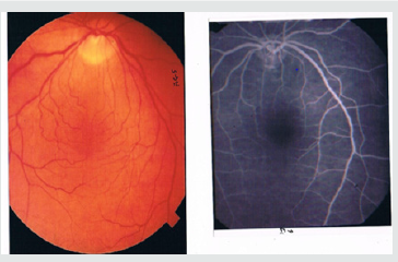
b) Resolution of Fundus Lesions: - The typical fundus picture of the cases is given which showed dilatation of the affected vein and venules with hemorrhages and macular edema in all the cases (Figure 2). The pre-treatment fluorescein angiography showed leakage of dye on the optic disc, in areas along the course of vein (Figure 3) and after the treatment, it showed restoration of blood flow with decrease in size of the venules, veins and absence of macular edema (Figure 4). The lesions started resolving after the day of treatment and completely resolved in 4-6 weeks (5 weeks).

c) Complication: - No complications had occurred till date [4]. There was no development of neo-vascularization in the retina, or in the angle of the anterior chamber. No case had developed thrombotic glaucoma.

Discussion

The idea of blasting of the dotted blood in the blocked central retinal vein struck to me from the use of Yag laser in removing the pigment dust from the surfaces of some implanted intra-ocular lenses. It was noticed that if we kept the settings of slit lamp at 125micron anterior and gave a burst of 1-1.5milli Joules, the dust got detached on a wider area of the implanted intra-ocular lens without damaging the lens [5,6]. This analogy was applied in this study on 2 blind eyes that showed complete safety of this modality, as there were no complications in the follow up, which further confirmed our idea to use it for this purpose. In this study, 10 cases were studied by this technique. Review of the literature showed that the initial improvement of the visual acuity with other procedures took about 1-4 months5:7’5 as a result the improvement seen after their procedures could not be definitely said to result from the intervention only [7]. The visual acuity of the patients was finger counting at 1meter distance prior to the treatment. However, in the laser clot blast therapy, the visual acuity improved to 0.4 (20/50) on 2nd day, which was clear cut proof of the beneficial effect of the technique. It appeared that the Yag laser energy had disintegrated the clotted blood in the blocked vein without damaging the vein or the adjacent retinal tissue. Some authors preferred to wait till complications of central retinal vein block started appearing. In our opinion, it was not correct but an absurd approach as the damage was bound to occur with uncertain outcome. Our study revealed that by opening of the flow in the blocked central vein at the earliest, not only improved the visual results but also shortened the recovery period with no subsequent complications. The treatment in the form of laser clot therapy should be carried out irrespective of the type of central retinal vein block. It should be carried out as an urgent outdoor procedure irrespective of the age, sex or associated disease. In the branch vein study groups, the final vision in laser treated eye improved to 20/40 to 20/50, compared with 20/70 in untreated eyes. Nevertheless, the treatment effect was negligible when the initial vision was in the poorer range of the 20/40 to 20/200 as inclusions criterion [8,9]. The treatment in the form of photocoagulation1 is destructive and is aimed to treat cases who develop neovascularization or non-resolving macular edema and not as an effort to correct the main problem of venous occlusion. The other surgical procedures in form of chorioretinal anastomosis, advential sheathotomy678 and cannulation° although seem logical but are potentially hazardous with indefinite outcome. The purpose of our investigation was an attempt to correct what we believed to be the pathogenic mechanism in central retinal vein occlusion. We tried to lyse the clotted blood or other intra-vascular factors, which had in our opinion, lead on to the stasis of venous blood [10]. This therapy proved useful as it improved the blood flow and abolished the occurrence of complications, which usually occur in this condition

 https://lupinepublishers.com/ophthalmology-journal/fulltext/laser-clot-blast-therapy-for-central-retinal-venous-occlusion.ID.000142.php

For more Lupine Publishers Open Access Journals Please visit our website: https://lupinepublishersgroup.com/

For more Trends in Ophthalmology Please Click

Here: https://lupinepublishers.com/ophthalmology-journal/

To Know more Open Access Publishers Click on Lupine Publishers 

Follow on Linkedin : https://www.linkedin.com/company/lupinepublishers
Follow on Twitter   :  https://twitter.com/lupine_online


Multiple Focal Choroidal Excavations in Association with Protein Rich Diet

  Introduction Choroidal excavation is a novel entity that is diagnosed with optical coherence tomography (OCT). In 1959, Klien,...