Friday, November 19, 2021

Lupine Publishers | Recent Changes in Tuberculosis Guidelines: What’s New for Ocular Tuberculosis

 Lupine Publishers | Trends in Ophthalmology Open Access Journal 
https://lupinepublishers.com/ophthalmology-journal/images/Lupinepublishers-openaccess-Ophthalmology.ID.000130.G001.png

Abstract

Ocular TB can cause moderate to severe visual impairment in up to 40% of affected eyes. Delay in diagnosis and treatment can result in chronic inflammation and loss of vision. The pathogenesis of ocular tuberculosis is still not clear, whether it result from a direct mycobacterial infection or a hypersensitivity response to mycobacterium tuberculosis. The choroidal nodules likely link from direct hematogenous infection while the vasculitis and choroiditis are probably the result of immune hypersensitivity. Prevalence of tuberculosis as an etiology of uveitis range from 5- 10% in endemic areas. Tuberculous uveitis inevitably leads to blindness if not properly diagnosed and treated. The aim of this paper is to review the presentation, diagnosis and management of tuberculous uveitis and to highlight the recent changes in the tuberculosis guidelines. Indian extrapulmonary tuberculosis (EPTB) guidelines were developed under the auspices of Central TB Division and Directorate of Health Services, Ministry of Health and Family Welfare, Government of India in 2016, with the objective of providing guidance on uniform, evidence-informed practices for suspecting, diagnosing and managing EPTB.

Keywords:ATT; Diagnosis; Ocular Tuberculosis; TST; Uveitis

Introduction

Tuberculosis (TB) is a global health problem. It is a chronic, granulomatous infection caused by Mycobacterium tuberculosis. This acid-fast bacillus usually affects the lungs but can affect any organ of body except teeth and hairs. Tuberculosis can be considered in the differential diagnosis of any type of intraocular inflammation. Because of the lack of standardized diagnostic criteria for tuberculous uveitis, and the difficulty of confirming the diagnosis by laboratory methods, epidemiologic data for tuberculous uveitis are unreliable. Incidence of tubercular uveitis in developed countries is less than developing countries. In India, where pulmonary TB is endemic, TB was the cause of uveitis in 5.6- 10.1% of the cases [1]. The aim of this review article is to focus on the diagnostic criteria and the management of tuberculous uveitis and to highlight the key changes as recommended in INDEX TB guidelines.

Pathogenesis

Ocular TB is either primary, in which the eye is the primary port of entry of the mycobacterium into the body, or secondary, as a result of seeding by hematogenous spread from a distant site. Primary disease usually involves eyelid, conjunctival, corneal, and scleral lesions, but are rare entities, while the secondary tuberculous disease involves uveal tract, retina, and optic nerve. Inflammation of the uveal tract is the most common eye manifestation of the disease, due to its high blood supply [2].

Diagnosis

Most patients with ocular involvement have no history of pulmonary or other systemic forms making a definitive diagnosis difficult [3-4]. Therefore, tuberculous uveitis is frequently under diagnosed and the disease is recognizable after a very long diagnostic delay [5]. The absence of clinically evident pulmonary TB does not rule out the possibility of ocular TB, as about 60% of patients with extra pulmonary TB have no evidence of pulmonary TB [6].

Ocular infection with M. tuberculosis is uncommon, but prevalence estimates may not be reliable because of diagnostic difficulties. Incidence of TB as a cause among patients presenting with uveitis has been reported at 10.1% in north India (Singh, 2004), but much lower in south India at 0.6% (Biswas, 1996-1997). This discrepancy may be due to several factors, including access to ophthalmology services, evolution of diagnostic criteria, description of new diagnostic entities and improvement in diagnostic tools [7].

There is always conflict between ophthalmologists and physicians for instituting anti-tubercular treatment (ATT) due to lack of gold standard criteria to diagnose ocular TB. Usually the patients referred to a Pulmonologist due to a suspicion of tuberculous uveitis will lack any clinical signs of a systemic disease on routine investigations and finally the decision to treat will lie on the ophthalmologist. The current practice at most of the centre is to make a presumptive diagnosis with positive TB skin test (TST), positive interferon gamma release assays (IGRA), and lesions on imaging of the chest (chest x-ray/ chest CT scan). Either resolution of uveitis or nonrecurrence of the uveitis after treatment completion of TB supports the diagnosis [7]. Before proceeding for full antituberculous treatment treating physician should always keep in mind the potential dangers of the therapeutic management.

There is a scarcity of data from clinical trials in EPTB and most of the information regarding diagnosis and management is extrapolated from pulmonary TB. Indian EPTB guidelines were developed under the auspices of Central TB Division and Directorate of Health Services, Ministry of Health and Family Welfare, Government of India in 2016, with the objective of providing guidance on uniform, evidence-informed practices for suspecting, diagnosing and managing EPTB.

The following are the key points to remember in case of ocular tuberculosis

Patients who require Ophthalmologist Review

Patients with symptoms consistent with anterior, intermediate, posterior or pan-uveitis, including the following: Red eye, blurred vision, photophobia, irregular pupil, eye pain, floaters, photopsia.

Investigations for Ocular TB

All suspected cases should undergo comprehensive examination including complete blood count, erythrocyte sedimentation rate, liver enzyme panel, infectious disease screen (which included Venereal Disease Research Laboratory (VDRL) test for syphilis, urine microscopy, serum ACE level), chest x ray, mantoux test, Quantiferon Gold test, and HRCT Chest. Blood should be taken for QTB Gold before the performance of mantoux test. PCR assays for TB DNA can be performed in patients with severe anterior chamber inflammation to confirm TB.

Ocular Imaging: Fundus photography, fluorescein angiography or optical coherence tomography to assess extent or complications of disease, and to monitor response to treatment.

X-ray of Chest and CT of the Chest: To find out the evidence of past or present MTB infection. A CXR with evidence of pulmonary nodules, calcification or fibrosis in the hilar area or upper lobes was considered as a positive CXR finding.

HRCT chest with evidence of parenchymal cavitating lesions, nodular lesions with tree in bud appearance, lymph node with center necrosis, matting and pleural effusion, hilar and mediastinal lymphadenopathy were considered as active TB. HRCT chest with evidence of calcified granulomas predominantly in upper lobes, calcified mediastinal lymph nodes and apical scarring were considered as healed TB.

Immunological Test: Tuberculin Skin Testing (TST)/ Interferon-Gamma Release Assays (IGRA) (Quantiferon Gold): Can be performed in all suspected cases. The predictive value of TST varies depending on the incidence of TB in the population and local BCG vaccination policy. IGRA is based on gamma interferon production by T cells sensitized to MTB specific antigens. It is not influenced by BCG and most nontuberculous mycobacteria (NTM). Positivity of both tests only indicates exposure to tuberculosis/ latent infection, and it is not a indicator of active TB disease. While a positive result may support the diagnosis of TB, a negative test cannot rule out TB.

CBNAAT Testing of Vitreous or Aqueous Specimens: Can be done in selected cases. The accuracy of Cartridge based nucleic acid amplification technique (CBNAAT) seems to vary significantly, but this is the only specific test that may identify ocular TB. Vitreous/ aqueous humour sampling by a trained person is recommended for CBNAAT testing.

Biopsy: Recommended in highly selected cases as biopsy of the structures of the eye is highly invasive and carries the risk of exacerbating visual loss. Specimens need histopathological examination with staining for acid-fast bacilli (AFB) and culture. So, should be performed in highly selected cases by the experienced persons.

Newer molecular methods like CBNAAT and line probe assay helps in diagnostic algorithm from any fluid/tissue samples for rapid detection and they also tells about drug sensitivity.

Diagnostic Categories

Can be divided into 4 categories

Presumptive Ocular TB: Patient with one of the following clinical presentations [7]:

I. Anterior Uveitis:

a. Granulomatous, mutton-fat keratic precipitates

b. Iris nodules

c. Broad-based posterior synechiae

d. Hypopyon may be observed

e. Vitritis

II. Intermediate uveitis

III. Posterior Uveitis: Subretinal abscess, multifocal choroiditis, choroidal/disc granuloma, multifocal serpiginous choroiditis and retinal periphlebitis.

IV. Panuveitis

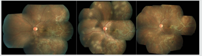
V. Rarely, scleritis (anterior and posterior), interstitial and disciform keratitis Figure 1.

Note: Extraocular TB disease is often absent in ocular TB patients and patients do not usually have systemic symptoms of fever and weight loss. Of all the clinical manifestations, the presence of choroidal granulomas, occlusive retinal vasculitis, and multifocal serpiginoid choroiditis has been more strongly associated with presumed ocular tuberculosis. In particular, choroidal granulomas should raise a high index of suspicion and failure to recognize them could lead to devastating consequences.

Possible Ocular TB:

a. At least one clinical sign suggestive of ocular TB with exclusion of other aetiologies.

b. Documented exposure to TB (history of contact with sputum positive pulmonary TB case) and/or immunological evidence of TB infection (TST/Quantiferon gold test positivity). Latent TB: Some of the patients had no evidence of active TB at another extraocular site. Demonstration of the presence of an immune response to TB antigens (Positive Mantoux or Quantiferon gold test) in the absence of evidence of active TB has been used to characterise the state of ‘latent’ TB. This term becomes confusing in the presence of active uveitis; this may be unrelated to the TB immune response, which therefore is truly latent. However, the uveitis may be an ophthalmic manifestation of TB, either directly or via a hypersensitivity mechanism. Despite the absence of other sites of active TB Ophthalmologist used to treat such cases as active TB.

Clinically Diagnosed Ocular TB (Probable Ocular TB):

a. At least one clinical sign suggestive of ocular TB with exclusion of other aetiologies.

b. Chest X-ray consistent with TB infection/Clinical evidence of extraocular TB/ microbiological confirmation from sputum or extraocular sites.

c. Documented exposure to TB (history of contact with sputum positive pulmonary TB case) and/or immunological evidence of TB infection (TST/Quantiferon gold test positivity).

Bacteriologically Confirmed Ocular TB: Microbiological (smear/culture) or, histopathological confirmation of MTB from ocular fluids/tissues.

Treatment of Ocular TB

Treatment with ATT with or without other adjuvant therapy is recommended in all patients with possible ocular TB, clinically diagnosed ocular TB or bacteriologically confirmed ocular TB, with the aim to protect visual function, to control ocular inflammation and to prevent recurrence of inflammation.

First Line Treatment for Adults and Children with Ocular TB: Principle of treatment of TB has been now daily regimen with administration of daily fixed dose combination of first line ATT as per appropriate weight bands for both newly diagnosed and previously treated patients. (Both the Category II and III has been discontinued now).

2(HRZE) + 4 (HRE): Continue ethambutol in continuation phase too (new guideline) (H-Isoniazide, R-Rifampicin, Z-Pyrazinamide, E-Ethambutol).

Duration of extrapulmonary TB treatment (new guideline) – The CP in both new and previously treated cases may be extended based on clinical decision of the treating physicians. Adjunctive treatments are corticosteroids (local or systemic) and other immunosuppressants.

I. Duration: Total treatment duration: 6 to 9 months.

II. Follow-up: Patients need regular monitoring during and after treatment with clinical evaluation (Slit-lamp biomicroscopy and Indirect Ophthalmoscopy). Fundus photo, fundus autofluorescence, fluorescine angiography and optical coherence tomography (OCT).

Follow-up visits requires initially monthly, then 2–3 monthly (or more frequently as required) to monitor response to therapy with a minimum period of 6 months after completion of therapy.

Treatment Outcomes: In ocular TB defined differently than other pulmonary and extrapulmonary tuberculosis, as microbiological confirmation of TB is rarely possible in ocular tissues. Thus, treatment success or failure is primarily guided by the level of inflammation seen inside the eye. The main outcome measure was recurrence of inflammation after minimum six months of initiating treatment.

Remission: Inactive disease for at least 3 months after discontinuing all therapy based on the Standardization of Uveitis Nomenclature recommendations (Jabs, 2005).

Treatment Failure: No decrease in inflammation, or less than a two-step decrease in level of inflammation after 3 months of ATT. Relapse: An increase in the level of inflammation after complete remission (at least two-step increase).

Approach to Treatment Failure

a. Work-up for other Etiology: Detailed ocular and systemic evaluation.

b. Look for Paradoxical Reaction: usually occurs within 2 months of starting ATT; responds to continuation or escalation of corticosteroid.

c. Rule out Drug Resistance: After ruling out first two points check history of contact with MDR TB patients and if facilities exist, consider ocular fluid sampling for molecular diagnosis of drug resistance.

d. Surgery: Is indicated in ocular TB in cases of complications of retinal vasculitis like retinal neovascularisation, vitreous haemorrhage or retinal detachment or complication of uveitis such as cataract and glaucoma.

Note: Introduction of Bedaquiline as a new drug. ATP synthase inhibitor specifically targets mycobacterial TB. Indicated in age more than 18 years [7].

Conclusion

Treatment with ATT with or without other adjuvant therapy is recommended in all patients with possible ocular TB, clinically diagnosed ocular TB or bacteriologically confirmed ocular TB. Principle of treatment of TB has been now daily regimen with administration of daily fixed dose combination of first line ATT for 6-9 months, as per appropriate weight bands for both newly diagnosed and previously treated patients, with continuation of ethambutol in continuation phase too. We recommend a high index of suspicion for tuberculous uveitis and a readiness to use ATT even where tuberculous aetiology has not been proven microbiologically but having clinical or radiologic findings of either active or latent TB infection and monitor for a response, in contrast to other forms of tuberculosis where this approach is not recommended. Randomized control trials are needed in the future but may be difficult to plan due to the low prevalence rate of ocular tuberculosis and until definitive diagnostic tests are available.

https://lupinepublishers.com/ophthalmology-journal/fulltext/recent-changes-in-tuberculosis-guidelines.ID.000130.php

For more Lupine Publishers Open Access Journals Please visit our website:

https://twitter.com/lupine_online

For more Trends in Ophthalmology Please Click

Here: https://lupinepublishers.com/ophthalmology-journal/

To Know more Open Access Publishers Click on Lupine Publishers 

Follow on Linkedin : https://www.linkedin.com/company/lupinepublishers
Follow on Twitter   :  https://twitter.com/lupine_online

Saturday, November 13, 2021

Lupine Publishers | The Mystery of Ectopia Lentis -A Rare Case Report

Lupine Publishers | Trends in Ophthalmology Open Access Journal 


Abstract

We report a case of a 12-year-old male with hematological, ocular, dermatological, dental and gastrointestinal involvement. To the best of our knowledge and after an extensive internet search, this probably is the first case of subluxation of the natural crystalline lens (NCL) with so many systemic associations described plus the pattern of subluxation itself.

Keywords: Ocular; Systemic; Associations

Case Report

A 12-year-old male (Figure 1) receiving treatment for gastroenteritis from the department of pediatrics (at our institute) presented to us with a history of itching bilateral eyes since his admission to the hospital. Secondly, the department of pediatrics diagnosed the child to be suffering from aplastic anemia and was referring him to a higher center for further management. On the contrary, old records bought by the patient’s father and mother showed that the patient was diagnosed as a case of thalassemia from some other institute and was receiving treatment in the form of regular blood transfusion from that institute. Further, the patient had multiple attacks of diarrhoea in the past few years for which he was admitted in hospital many times. The diarrhoea was not related to any particular meal. He was also receiving treatment from dental department for grade 3 mobility of lower anterior teeth. The positive findings on his general physical and systemic examination were thin sparse hairs on the scalp (Figure 2), facial freckles (Figure 3), and barrel shaped chest (Figure 4). His blood sugar, renal function tests, liver function tests, X ray chest, electrocardiography, vitamin B12 plus folate and zinc levels, C reactive proteins and serum electrolytes which were already carried out were within normal limits. The patient’s parents were farmers by occupation and did not give a history of themselves suffering from any major illness in the past. The patient’s family history revealed that one of the siblings had died at a younger age because of some illness, though no detail or hospital records of the disease were available with the parents. There was no other significant medical, surgical, traumatic or drug usage history.

Ocular examination was carried out. His visual acquity was finger counting at 2 meters bilaterally with no improvement on pinhole and refraction. Slit lamp examination of the right eye (Figure 5) revealed inferonasal subluxation of the NCL with a shallow anterior chamber while the left eye (Figure 6) had subluxation of the NCL into the anterior chamber with a shallow anterior chamber. The intraocular pressure and axial lengths of both the eyes were within normal limits. The patient did allow us to do gonioscopy. Bilateral fundus could not be seen because of media haze, though the NCL bilaterally were not cataractous nor was there any intraocular inflammation. Bilaterally, the ocular movements and pupillary reaction were normal. The itching in his eyes was as a result of allergic conjunctivitis for which we prescribed him antihistaminic eye drops. Clinical features and investigations for diagnosis of homocysteinuria, marfans syndrome and ehler danlos syndrome did not reveal any abnormality. Facility for ultrasound biomicroscopy (UBM), Optical coherence tomography (OCT), B scan ultrasonography and genetic studies were not available at our institute. We could have planned for his NCL removal and a probable intraocular lens implantation under guarded visual prognosis, but the patient wanted all his treatment modalities to be done in single institution, and his referral center had all the facilities.

Discussion

Ectopia lentis is displacement of the lens from its normal position. Ocular causes of lens subluxation include trauma, high myopia, buphthalmos, anterior uveal tumors, pseudoexfoliation syndrome, and hypermature cataracts [1]. Systemic disorders associated with ectopia lentis include Marfan syndrome, homocystinuria, Weill-Marchesani syndrome, and Ehlers-Danlos syndrome [2].

Spontaneous dislocation of the NCL is an extremely rare entity, especially its dislocation to the anterior chamber. This dislocation to the anterior chamber can lead to various complications like corneal edema, pupillary block glaucoma and anterior uveitis which requires urgent removal of the NCL [3]. Surgical procedures such as intracapsular cataract extraction, limbal or pars plana lensectomy and anterior vitrectomy and suturing of the haptics to the sclera have been the surgical modalities in use. Femtosecond laser assisted cataract surgery, glued intraocular lenses, instead of conventional scleral fixation of IOLs with sutures for fixation are another useful advancement in treatment [4].

Conclusion

The presence of ectopia lentis with multisystem involvement in our patient could be a part of a syndrome complex or may be an isolated entity it still remains a mystery for us. Ectopia lentis with hematological involvement has probably not been reported before (to the best of our knowledge)

https://lupinepublishers.com/ophthalmology-journal/fulltext/the-mystery-of-ectopia-lentis-a-rare-case-report.ID.000129.php

For more Lupine Publishers Open Access Journals Please visit our website:

https://twitter.com/lupine_online


For more Trends in Ophthalmology Please Click

Here: https://lupinepublishers.com/ophthalmology-journal/

To Know more Open Access Publishers Click on Lupine Publishers 

Follow on Linkedin : https://www.linkedin.com/company/lupinepublishers
Follow on Twitter   :  https://twitter.com/lupine_online

Tuesday, November 2, 2021

Lupine Publishers | A Case and Cranial MRI Characteristics of Progressive Supranuclear Palsy

 Lupine Publishers | Trends in Ophthalmology Open Access Journal 


Abstract

Progressive supranuclear palsy (PSP) is a neurodegenerative disease described by vertical supranuclear gaze paralysis, axial rigidity and postural instability involving falls in the early stage of the disease. Imaging studies present brain atrophy in especially midbrain, premotor cortex, basal ganglia and thalamus. There is no bioindicator of the disease although it is clinically identified. PSP is also classified as tauopathy. However, no specific indicator for tau protein could be identified. Hence, imaging findings, especially midbrain atrophy, functions as a biomarker in clinical practice and determination of disease prognosis. It has been shown that midbrain atrophy has a diagnostic value for PSP accompanied with the finding of “hummingbird” or “penguin” encountered by magnetic resonance imaging (MRI).

In this case report, we have aimed to review the diagnostic features of the disease based on the results of clinical and radiological results and to demonstrate the “penguin” sign and midbrain atrophy as a biomarker for clinical follow-up process. We have also detected a correlation with the results of clinical and neuropsychological tests.

We have analyzed a 61-year-old male patient who was a retired teacher for the complaints of progressive difficulty in walking since the recent year, slowness in speech fluency, forgetfulness and easy falling. Our patient was performed 1.5 Tesla Cranial MRI study due to the signs of postural instability, easy falling backward while walking, cognitive impairment, apathia, conjugate vertical gaze palsy, disinhibition and anxiety. T1W, T2W, T2 flair and diffusion sections on the axial plane were obtained in this imaging study. In addition, Mini-Mental State Examination and neuropsychological test were also performed in our patient.

The patient had particularly diffuse cerebral atrophy in predominantly frontal and parietal lobes. Peripheral CSF spaces were enlarged due to atrophy. Brainstem also showed atrophy and particularly mesencephalon atrophy was more remarkable. The appearance of brainstem in the sagittal slices due to mesencephalon atrophy were termed as “penguin” sign. A mild atrophy was also encountered in the bilateral cerebellar hemispheres. As a result of neuropsychological evaluation, mild to moderate cognitive impairment were detected.

We have evaluated with this case that atrophy of midbrain and mesencephalon which presented a significant visuality with classic “penguin” sign has an important diagnostic value in the PSP cases. Additionally, we have reviewed the fact in the light of literature data that imaging of midbrain atrophy may play a role as a biomarker in performing clinical follow-up and determination of the prognostic features of the PSP cases.

Introduction

Progressive supranuclear palsy (PSP) is a neurodegenerative disorder of primitive tauopathies [1]. It is characterized with pseudobulbar paralysis, dysarthria, dystonic rigidity of neck and trunk, akinesia, postural instability involving frequent falls and frontotemporal dementia as well as vertical gaze paralysis [2]. PSP is the most commonly seen atypical Parkinsonism. Its prevalence is 6.0-6.4 in 100,000. Its incidence is 5.4 cases in 100,000 [3]. It should be differentiated from idiopathic Parkinson’s disease or other atypical Parkinsonism cases along its natural clinical course [4,5]. The neuropathologically determinant characteristic of PSP is midbrain atrophy.

Atrophy is associated with intensity of tau-positive aggregates which are not melted in any way. These tau-positive formations are neuronal and glial fibrillar tangles and neurophil threads [6- 8]. This abnormal accumulation of tau proteins are responsible for the neuronal degeneration of basal ganglia and midbrain nuclei. Midbrain atrophy involves tegmentum, tectum, quadrigeminal layer and periaqueductal gray matter and is associated with reduced intensity of myelinated fibers, gliosis and neuronal loss. It is more commonly observed in males. Retropulsion and postural instability observed at early stage lead to frequent falls. The occurrence of the falls within the initial onset year is a typical characteristic. On the other side, supranuclear gaze paralysis usually appears due to primarily slowing of vertical saccades. Dystonia in the frontalis and procerus muscles may give a facial expression of scared or surprised in the PSP patient. Different clinical types of PSP have been recently identified [6]. In PSP patients, especially attention, executive function loss, verbal and nonverbal memory deficiencies may develop. The performance of the patient is demonstrated by verbal fluency tests, Frontal Assessment Battery and Montreal Cognitive Assessment Battery [4,9].

Case

The 61-year-old male patient was evaluated for the complaints of difficulties in starting walking since the recent year, frequent backward falls while walking, slowed speech and eye opening apraxia. He was conscious however, he had slow speech fluency and hypophonic dysarthria. His blink reflex was reduced. He had upward and downward supranuclear gaze paralysis and axial rigidity. He was able to walk independently with unsafe wide steps in the short distance. “Pull” test result was positive. His emotional state was labile and a remarkable slowing started in the executive functions. No response to the treatment of L-Dopa could be obtained.

Methods

The case was performed 1.5 Tesla MRI study. Cranial MRI was performed in accordance with T1weighted axial plane (slice thickness:5.5mm;TR:560;TE:9.5, Matrix 288x182), T2weighted sagittal plane (slice thickness: 5mm, TR: 5300, TE:82, Matrix: 384x192), T2weighted flair on the axial plane (slice thickness:5.5 mm,TR:8000, TE: 98, TI:2000, Matrix:320x224); T2weighted axial plane (slice thickness: 5.5mm,TR:3700, TE:88 Matrix: 320x256) (Figure 1). In addition, Montreal Cognitive Assessment Battery (MOCA Test) was applied. “Benton” facial recognition test and Line Direction Tests were performed.


Discussion and Conclusions

PSP is a clinical diagnosis. There is no diagnostic laboratory test. Midbrain atrophy that is frequently encountered by Cranial MRI may play a role as biomarker in determination of the prognosis. Especially, image of “penguin” encountered in the sagittal MRI slices has a diagnostic value. The progression of Parkinson’s Disease which is one of the diseases to be considered in differential diagnosis is very much slower than PSP and has no “red flag symptoms” and its responsiveness to L-Dopa is very well [1-4].

Corticobasal degeneration is a tauopathy like PSP, however, it is characterized with Parkinsonism manifested by remarkable symmetric dystonia. It is accompanied by cortical sensory signs, progressive apraxia is typically observed and “Alien Hand Syndrome” occasionally appears. It is differentiated by autonomic involvements such as Multiple System Atrophy with orthostatic hypotension, cerebellar ataxia and inspiratory stridor. Parkinson’s-Dementia complex manifested by Lewy body dementia and Parkinsonism is differentiated by fluctuating mental state and visual hallucinations appearing with onset of dopaminergic treatment [1-6].

Cranial MRI of the case encountered diffuse cerebral atrophy in predominantly frontal and parietal lobes. Peripheral cerebrospinal fluid (CSF) spaces were enlarged due to atrophy. Atrophy was present also in the brainstem and the atrophy particularly in the mesencephalon is much more remarkable. The appearance of the brainstem due to mesencephalon atrophy in the sagittal slices was termed as “penguin sign”. Additionally, bilateral cerebellar hemispheres revealed a slight atrophy [7-9].

According to MOCA Test scores, a significant decline was monitored in the copying and visuospatial functions of the patient. The patient gained no point from the subfield of executive functions. He has experienced difficulties in ordering the parameters in a correct sequence in this subfield. He could not order the parameters (1-5, A-E) as a number followed by a letter although he was a retired teacher. This result indicated a significant problem in the executive functions of this patient. In this subfield, the patient could not copy a cube and could gain no point from the subfield of clock-drawing test (Figures 2 & 3). The patient gained 5 standard deviations lower than the mean of his age and educational level according to the result of Benton Test of Facial Recognition. He was found significantly unsuccessful in Line Direction Tests. Evaluation of visual performance tests: It was observed that the patient’s cognitive processes related to orientation, spatial perception, spatial cognition, mental rotation, and face recognition were significantly impaired

https://lupinepublishers.com/ophthalmology-journal/fulltext/a-case-and-cranial-mri-characteristics-of-progressive-supranuclear-palsy.ID.000128.php 

For more Lupine Publishers Open Access Journals Please visit our website: https://twitter.com/lupine_online

For more Trends in Ophthalmology Please Click

Here: https://lupinepublishers.com/ophthalmology-journal/

To Know more Open Access Publishers Click on Lupine Publishers 

Follow on Linkedin : https://www.linkedin.com/company/lupinepublishers
Follow on Twitter   :  https://twitter.com/lupine_online


Multiple Focal Choroidal Excavations in Association with Protein Rich Diet

  Introduction Choroidal excavation is a novel entity that is diagnosed with optical coherence tomography (OCT). In 1959, Klien,...